A New Tool for Reviewing Dental Images
Dental images help dentists assess tooth decay, including areas that may be difficult to inspect during a visual examination. Artificial intelligence, or AI, is a developing tool that can analyze images for patterns and flag places that may deserve closer attention. It does not think or interpret images in the same way a dentist does, and its findings still need professional review. An overview of AI in dentistry describes the technology as a way to assist with tasks that involve pattern recognition and problem-solving.
Research on AI-assisted caries detection is promising, but results vary across systems and image types. A 2025 review found pooled sensitivity of 0.85 and specificity of 0.90 across the studies included in its analysis, but those estimates are not guarantees for a particular patient or practice. The review assessed diagnostic accuracy, not whether AI consistently speeds diagnosis in everyday dental care. The review’s findings support viewing AI as an aid, not a replacement for a dentist’s assessment.
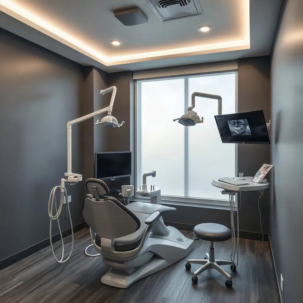
At V Dental in Live Oak, digital X-rays are among the technologies used to support dental care, alongside CBCT imaging, intraoral cameras, and iTero scanning. The practice information provided does not state that V Dental uses an AI diagnostic system. Patients can ask their dentist how their images are reviewed and how any finding fits with an examination and personal treatment plan.
How AI Flags Possible Tooth Decay
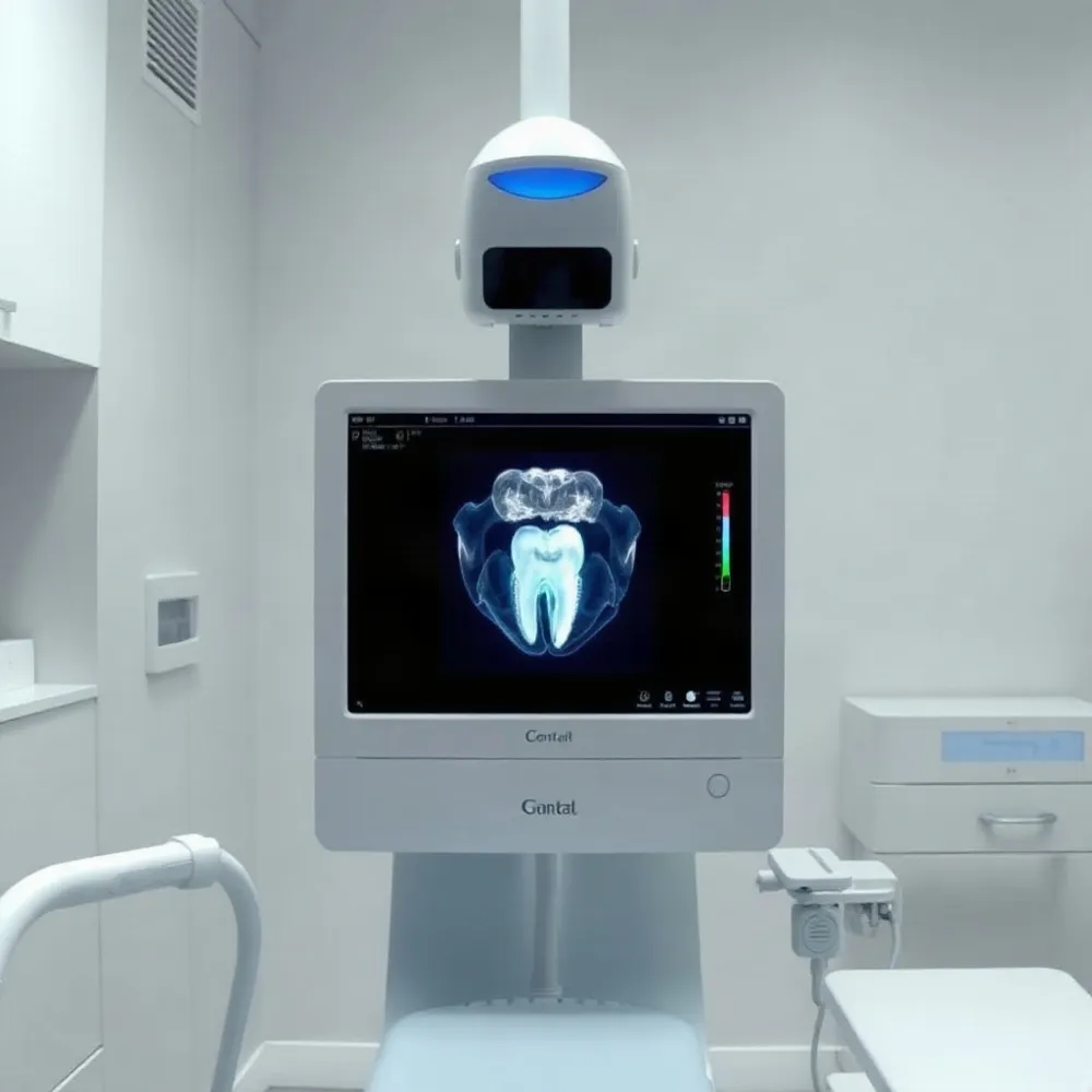 Computer-vision systems are trained on dental images to recognize patterns that may be associated with tooth decay. When a system detects a possible concern, it can flag the area for a dentist to inspect more closely. Research reviews describe this image analysis as support for clinical decision-making, not an independent diagnosis (scoping review of AI image analysis in dentistry).
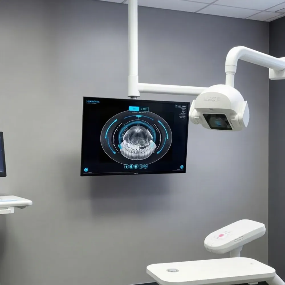
Some tools add visual annotations or overlays to an X-ray, drawing attention to a suspected finding. The way alerts appear varies by product, so a highlight should be understood as a prompt for review, not proof that decay is present. It may help focus attention on a part of the image that warrants a closer look.
At V Dental, digital X-rays are part of a broader imaging toolkit that also includes CBCT 3D imaging and intraoral cameras. These tools provide images for a dentist to assess, while any AI suggestion, where available, would need to be checked against the image and the patient’s clinical circumstances.
To decide whether a flagged area represents caries, the dentist considers the image alongside a clinical examination, symptoms, and dental history. The dentist may agree with an alert, dismiss it, or seek additional information before recommending care. Research guidance emphasizes that practitioners remain responsible for diagnosis and clinical judgment (AI and dental caries detection).
A marked image can also give patients a clear visual reference during a conversation about a finding and possible next steps. The dentist can explain what the area may indicate, what remains uncertain, and whether monitoring, preventive care, or treatment is appropriate. That discussion keeps the image in context rather than treating a software alert as a final answer.
What Accuracy Studies Tell Us
 How accurate and reliable is AI at identifying cavities? A 2025 umbrella review and meta-analysis offers a useful estimate, while also showing why no single score can guarantee what a system will find in a particular patient.
The researchers examined 14 systematic reviews and identified 137 original studies. Only 20 reported enough diagnostic information to be included in the quantitative analysis, which covered 29,423 diagnostic tests. The review reported pooled sensitivity of 0.85, specificity of 0.90, and an area under the receiver operating characteristic curve (AUC) of about 0.86 in its analysis.
Sensitivity describes how often a test correctly identifies images that show caries. Specificity describes how often it correctly distinguishes images without caries. These pooled results suggest that AI could help identify patterns in dental images, but they are research estimates across varied studies, not guarantees for every patient, product, or practice.
The review also estimated a 79% probability of caries after a positive AI result and a 6% probability after a negative result. Those figures depended on the study data, including a caries prevalence of 27.3% among the analyzed tests, so they should not be treated as universal predictions for an individual.
Performance varied substantially between studies. Imaging type, software design, training data, and the population being assessed can all affect results. AI may miss decay or flag an area that is not caries, and the review does not show that AI consistently matches or outperforms individual dentists.
At V Dental, digital X-rays and CBCT 3D imaging are part of a broader diagnostic approach. Any image finding still needs to be considered alongside a dentist’s examination, symptoms, and history. The evidence supports treating AI results as one source of information, not as a standalone diagnosis.
Images and Lesions Affect Performance
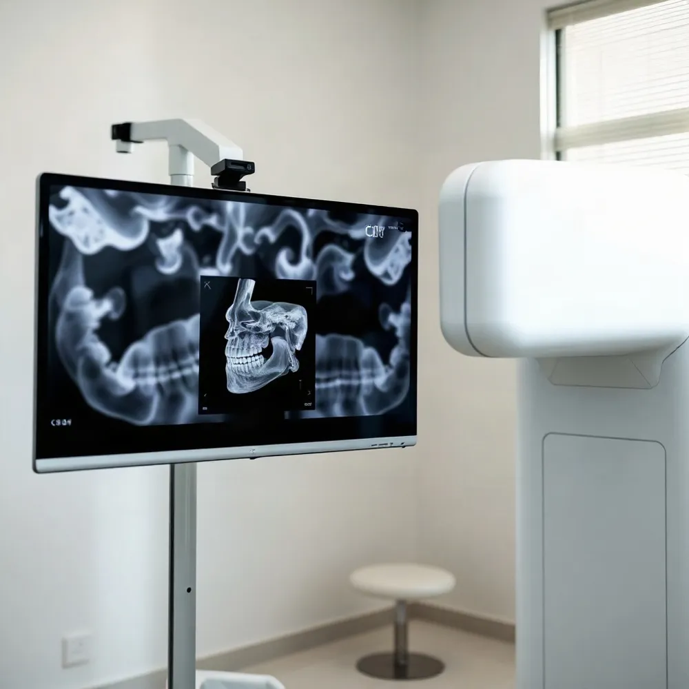 An AI system’s results depend partly on the kind of image it analyzes and the lesion it is asked to detect. Caries research has used bitewing, periapical, and panoramic radiographs, CBCT images, intraoral photographs, and near-infrared transillumination. Performance measured with one image type cannot automatically be assumed for another, as a 2025 umbrella review of AI caries detection cautions.
Image features also matter. Bitewings can show surfaces between neighboring teeth clearly, while panoramic images may have overlapping teeth that make some areas harder to assess. V Dental uses digital X-rays and CBCT imaging as part of its diagnostic technology, but findings from research on a particular image type do not establish how an AI tool would perform in that practice.
A study of 50,179 intraoral tooth photographs reported different results for front and back teeth, and for early white-spot lesions versus cavitated decay. For example, the model’s reported sensitivity and specificity varied across those groups, showing why one headline performance figure can hide meaningful differences. The study does not establish that the same results apply to every patient or dental practice (study indexed by PubMed).
A separate study focused on decay beneath restorations or around braces. It analyzed 505 bitewing radiographs and reported promising results for its models, while calling for larger, more varied datasets and validation in real clinical settings (2025 study in Bioengineering). The researchers also noted that cervical burnout and some radiolucent restorative materials can resemble caries on an X-ray, potentially leading to false positives.
V Dental also uses intraoral cameras, which provide another view of the teeth. Whatever the image source, a highlighted area is a prompt for the dentist to assess, not confirmation of a cavity on its own.
Why Results Need Clinical Context
A result from an AI image review is only as useful as the evidence behind the system and the fit between its training data and the patient in front of the dentist. The 2025 umbrella review found substantial variation across studies, so its pooled estimates should not be treated as a guarantee for every image or practice.
The evidence base also had gaps. Of 137 original studies identified, just 20 reported the diagnostic counts needed for the meta-analysis, and the authors found signs of publication bias. The analyzed tests had an estimated caries prevalence of 27.3%, which the review said was lower than prevalence in the community. That difference raises questions about how well some study datasets represent patients seen in routine care. The umbrella review recommends broader representation across populations and healthcare settings.
Many studies also did not report lesion depth or location in enough detail to show whether performance changes for early versus deeper decay, or for one tooth surface versus another. Researchers called for more focused study of lesion site and depth, along with validation under real-world clinical conditions.
For patients at V Dental, digital X-rays and CBCT imaging form part of a wider assessment that can include an examination and discussion of symptoms and history. An AI-highlighted area should be treated as a prompt for the dentist to evaluate, not proof of a cavity or an automatic reason to recommend treatment. The dentist weighs the image with the rest of the clinical picture before discussing next steps.
AI Supports the Dentist, Not Replaces Them
An AI system can flag an area that may show decay, but a flag is not a diagnosis. A 2025 umbrella review concluded that AI can assist with caries detection while leaving diagnosis and responsibility with dental practitioners review of AI caries-detection accuracy.
Dentists interpret image findings alongside a visual examination, symptoms, dental and medical history, and the patient’s individual circumstances. A shadow on an X-ray may warrant a closer look, but the dentist must decide whether it reflects decay and what, if anything, should be done. AI is a decision-support aid, not a substitute for professional judgment discussion of AI’s role in dentistry.
A system may provide an additional, consistent review and draw attention to a spot that deserves inspection. That can help organize the dentist’s assessment, but the available review does not establish that AI routinely shortens appointments or speeds diagnosis in everyday practice. Research performance should not be treated as a promise about an individual patient’s result.
Safe use also depends on suitable training data, privacy and security safeguards, professional training, and human oversight. If the images or patient groups used to develop a system differ from those seen in practice, its output may be less relevant. Dentists need to understand a tool’s limits and check its suggestions against the clinical picture.
V Dental’s stated technology includes digital X-rays, CBCT 3D imaging, intraoral cameras, and iTero scanning. These tools support image capture and assessment, but their use does not establish that the clinic offers AI image analysis. Patients can ask their dentist how any finding was assessed and what evidence supports a recommended next step.
A Helpful Signal, Not a Final Answer
An AI flag can draw attention to an area that may show decay, giving a dentist another prompt to review the image. It cannot guarantee that a cavity is present, rule one out with certainty, or promise a faster diagnosis for every patient. The result is one piece of evidence, not a treatment decision.
If an image raises a concern, ask your dentist what the finding may mean and how it fits with the clinical examination, symptoms, and dental history. A dentist can explain whether monitoring, preventive care, or treatment is appropriate for your situation. At V Dental, digital X-rays are one part of a broader approach that includes personalized treatment plans, rather than decisions based on an image alone.
Research suggests AI may assist with caries detection, but findings across systems and imaging types are not uniform. Broader clinical validation, using diverse patient groups and real-world settings, will help show where these tools can contribute reliably. Until that evidence is clearer, patients can treat an AI-generated flag as a reason for a conversation with their dentist, not as a diagnosis or a recommendation to undergo treatment.